

Oxford Biomedica
Single-step nuclease treatment in lentiviral manufacturing
About tenfold lower total DNA and threefold lower plasmid DNA than the standard nuclease process, while removing the second nuclease step.
Up to 70% of produced viral vector is lost in downstream processing, and host-cell chromatin left by conventional nucleases is a hidden driver of that loss.
Lentiviral and other fragile enveloped vectors, including oncolytic and vaccine viruses such as measles and enveloped VLPs, need a nuclease that works in cell media. Matching it to these condition scan up to double recovery and cut modeled cost per dose by about 40%.
Up to 70% of produced viral vector is lost in downstream processing, and host-cell chromatin left by conventional nucleases is a hidden driver of that loss.
Lentiviral and other fragile enveloped vectors, including oncolytic and vaccine viruses such as measles and enveloped VLPs, need a nuclease that works in cell media. Matching it to these condition scan up to double recovery and cut modeled cost per dose by about 40%.
In lentiviral and other enveloped virus production, reducing residual host-cell DNA is a critical part of downstream processing. But not all host-cell DNA is equally accessible to nuclease treatment.
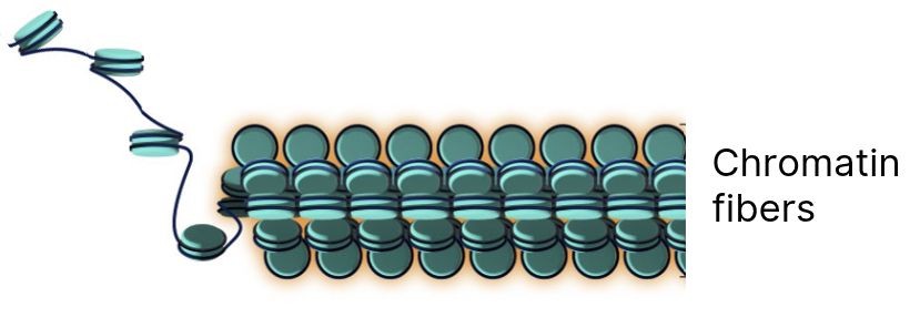
A significant proportion of host-cell DNA can remain associated with histones as chromatin, forming compact nucleoprotein structures. This limits nuclease access and makes chromatin-associated DNA more difficult to digest than free DNA. Under process conditions, the nuclease therefore has to break down not only accessible DNA, but DNA protected within chromatin.
Chromatin accessibility is only part of the challenge. Nuclease activity also depends on the conditions in which digestion takes place.
Conventional nucleases from S. marcescens lose more than half of their activity at physiological salt. Lentiviral supernatants typically sit at 130–155 mM NaCl (the physiological range in Figure 2) and near-neutral pH (7.2–7.4), sometimes with a little more salt added. That is already well outside the optimal range of conventional nucleases, so chromatin digestion stays incomplete. Raising salt to compensate stresses the envelope, and the usual workarounds each add cost: more nuclease, an extra desalting step, or a second nuclease treatment after chromatography.
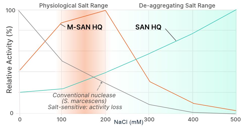
Fig 2. Relative activity of nucleases across salt concentrations
Conventional nucleases (S. marcescens) lose activity as salt rises. Salt-adapted nucleases keep it: M-SAN HQ in the physiological salt range of cell culture media, SAN HQ in the de-aggregating salt range.
ArcticZymes testing conditions →Chromatin fragments that survive nuclease treatment can persist through downstream processing alongside virus particles, with effects across the workflow:
Effective chromatin clearance therefore matters not only for downstream process performance, but also for controlling residual DNA.
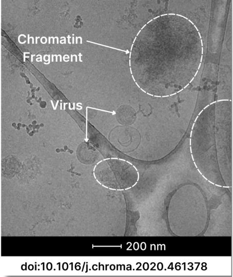
Fig 3. Chromatin fragments persist through conventional nuclease treatment and downstream processing, co-localizing with virus particles.
Image courtesy acib. Adapted from Pereira Aguilar et al., J. Chromatogr. A 1627 (2020) 461378.
View publication →
In lentiviral and other enveloped virus production, reducing residual host-cell DNA is a critical part of downstream processing. But not all host-cell DNA is equally accessible to nuclease treatment.
A significant proportion of host-cell DNA can remain associated with histones as chromatin, forming compact nucleoprotein structures. This limits nuclease access and makes chromatin-associated DNA more difficult to digest than free DNA. Under process conditions, the nuclease therefore has to break down not only accessible DNA, but DNA protected within chromatin.
Chromatin accessibility is only part of the challenge. Nuclease activity also depends on the conditions in which digestion takes place.
Conventional nucleases from S. marcescens lose more than half of their activity at physiological salt. Lentiviral supernatants typically sit at 130–155 mM NaCl (the physiological range in Figure 2) and near-neutral pH (7.2–7.4), sometimes with a little more salt added. That is already well outside the optimal range of conventional nucleases, so chromatin digestion stays incomplete. Raising salt to compensate stresses the envelope, and the usual workarounds each add cost: more nuclease, an extra desalting step, or a second nuclease treatment after chromatography.
Fig 2. Relative activity of nucleases across salt concentrations
Conventional nucleases (S. marcescens) lose activity as salt rises. Salt-adapted nucleases keep it: M-SAN HQ in the physiological salt range of cell culture media, SAN HQ in the de-aggregating salt range.
ArcticZymes testing conditions →Chromatin fragments that survive nuclease treatment can persist through downstream processing alongside virus particles, with effects across the workflow:
Effective chromatin clearance therefore matters not only for downstream process performance, but also for controlling residual DNA.
Fig 3. Chromatin fragments persist through conventional nuclease treatment and downstream processing, co-localizing with virus particles.
Image courtesy acib. Adapted from Pereira Aguilar et al., J. Chromatogr. A 1627 (2020) 461378.
View publication →
In lentiviral and other enveloped virus production, reducing residual host-cell DNA is a critical part of downstream processing. But not all host-cell DNA is equally accessible to nuclease treatment.
A significant proportion of host-cell DNA can remain associated with histones as chromatin, forming compact nucleoprotein structures. This limits nuclease access and makes chromatin-associated DNA more difficult to digest than free DNA. Under process conditions, the nuclease therefore has to break down not only accessible DNA, but DNA protected within chromatin.
Chromatin accessibility is only part of the challenge. Nuclease activity also depends on the conditions in which digestion takes place.
Conventional nucleases from S. marcescens lose more than half of their activity at physiological salt. Lentiviral supernatants typically sit at 130–155 mM NaCl (the physiological range in Figure 2) and near-neutral pH (7.2–7.4), sometimes with a little more salt added. That is already well outside the optimal range of conventional nucleases, so chromatin digestion stays incomplete. Raising salt to compensate stresses the envelope, and the usual workarounds each add cost: more nuclease, an extra desalting step, or a second nuclease treatment after chromatography.
Fig 2. Relative activity of nucleases across salt concentrations
Conventional nucleases (S. marcescens) lose activity as salt rises. Salt-adapted nucleases keep it: M-SAN HQ in the physiological salt range of cell culture media, SAN HQ in the de-aggregating salt range.
ArcticZymes testing conditions →Chromatin fragments that survive nuclease treatment can persist through downstream processing alongside virus particles, with effects across the workflow:
Effective chromatin clearance therefore matters not only for downstream process performance, but also for controlling residual DNA.
Fig 3. Chromatin fragments persist through conventional nuclease treatment and downstream processing, co-localizing with virus particles.
Image courtesy acib. Adapted from Pereira Aguilar et al., J. Chromatogr. A 1627 (2020) 461378.
View publication →Salt-active nucleases keep their activity under process-relevant salt conditions, so they digest chromatin where conventional nucleases stall. For fragile enveloped vectors, that means anuclease built for physiological salt, working directly in cell media.
Researchers at acib and BOKU Vienna compared nucleases ahead of flow-through chromatography of measles virus, an enveloped virus. After treatment with M-SAN HQ, no chromatin was detected in the flow-through, while the viral nucleoprotein was preserved. With two conventional nucleases, a clear chromatin band remained (see the data - Mayer et al., Biotechnology Progress (2023), doi:10.1002/btpr.3342).

When compared with non-salt-tolerant endonucleases (Benzonase® and DENARASE®), salt-tolerant endonucleases have better performance at the salt concentrations and pH naturally present in the cell culture supernatant, allowing its direct application without further processing. In addition to that, salt-tolerant endonucleases allowed the removal of chromatin, one of the most critical impurities and often overlooked in enveloped virus DSP.
Activity under the process's own salt conditions decides whether chromatin is removed or becomes a filtration liability. Size is the reason: chromatin fragments range up to around 300 nm, overlapping the size of a lentivirus (80–100 nm) or measles virus (120–250 nm). For flow-through chromatography, the nuclease has to cut them below about 50 nm so they enter the resin while the virus passes through.
A conventional nuclease that loses activity at physiological salt leaves fragments in that range, so they are retained alongside the vector during size-based separation. A salt-active nuclease that stays active under process conditions cuts chromatin well below vector size, so the fragments are removedwhile the vector passes through.
There is no single right step for nuclease treatment, as long as the process stream stays at physiological salt:
A starting point for evaluation. If you start from clarified or partly purified material, begin at step 2.
Tip: if clearance is incomplete, check Mg²⁺ and pH before increasing the dose.
In a modeled 2,000 L lentiviralprocess, matching the nuclease to process conditions cut nuclease cost perbatch by more than 70% and cost per dose by about 40%. Download the whitepaper for the full model, the AAV and lentiviral scenarios, and the independentdata behind them.
Standard DNA assays measure accessible DNA, not the chromatin structures that drive filter fouling, aggregation and recovery loss. Chromatin is less accessible to dye-based assays such as PicoGreen, so disrupting it first, for example with proteinase K, often reveals significantly more residual host-cell DNA. That is also why downstream performance can improve even when a DNA readout barely changes. Find out more in our webinar: Your Viral Vector Analytics May Not Reflect Process Reality.
M-SAN HQ is made in Norway under an ISO13485:2016 quality management system, from raw materials of certified non-animal origin. It is an ancillary material and is not intended to be part of the final drug product.

“In our viral vector manufacturing processes, ArcticZymes’ M-SAN HQ and SAN HQ enzymes consistently deliver efficient host-cell DNA clearance under both physiological and high-salt conditions. This has allowed us to achieve reliable digestion with fewer nuclease units, improving process efficiency without compromising quality.”
Team Lead, Viral Vector Process Development, Revvity