
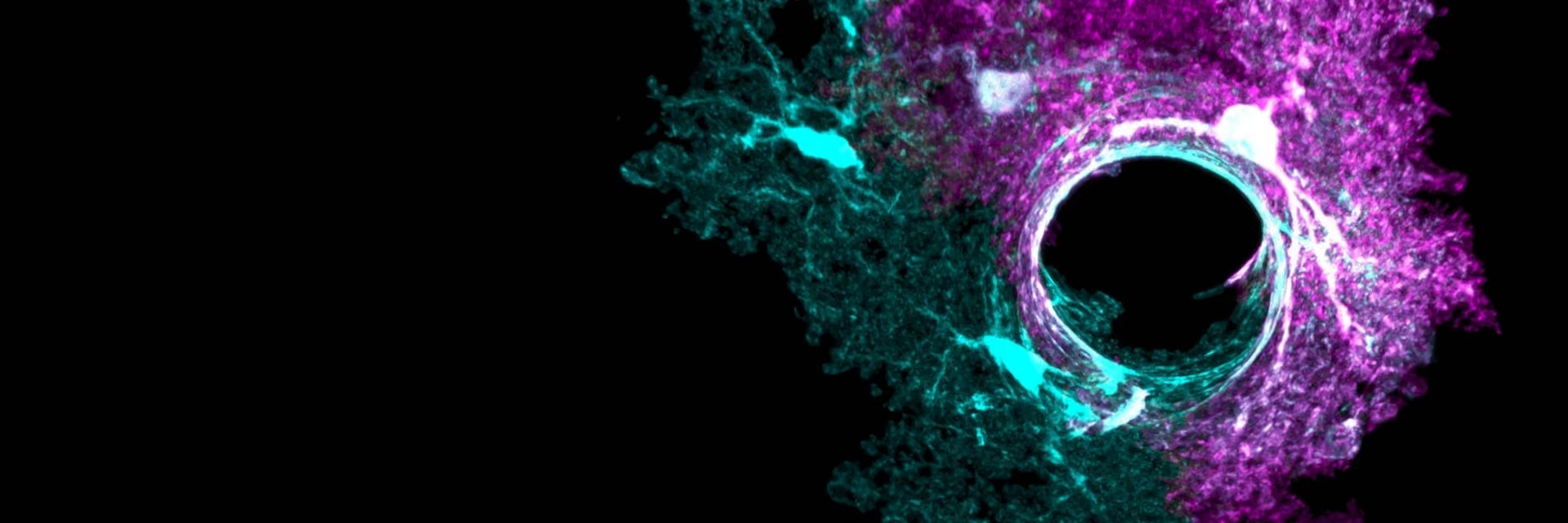
Adeno-associated viruses (AAVs) are widely used to deliver genetic material into specific cell types, but their quality, particularly specificity and purity, can significantly affect experimental results.
New tools developed at UCLA are giving researchers an unprecedented view of how brain cells respond to stroke, demonstrating the importance and capabilities of high-quality viral vectors.

Understanding the brain’s response to stroke
When someone has a stroke, the brain does not simply shutdown in the affected area. The surrounding tissue mounts an active response, with different cell types mobilizing to limit damage and, in some cases, begin rebuilding. One of the key players in this process is a type of cell called an astrocyte — a star-shaped, non-neuronal cell found throughout the brain that has long been thought of mainly as structural support, but is now understood to play a much more active role in regulating brain health and repair.
The biological question is straightforward enough: what are astrocytes actually doing after a stroke, and could they be nudged towards better repair? What held the field back was a technical problem that came from an unexpected direction — the tools available to study astrocytes were not specific enough.
The challenge of AAV specificity and off-target expression
Astrocytes sit alongside neurons throughout the brain, making cell-specific targeting with AAVs challenging. Many AAVs designed to target astrocytes also express their cargo in neurons, leading to off-target expression that directly impacts AAV quality.
That is a problem, because neurons and astrocytes play different roles, and if both cell types are affected, it becomes difficult to attribute any observed effect to a specific cell type.
Dr Amy Gleichman tackled this directly during her postdoctoral work at UCLA, working in the lab of Dr. S. Thomas Carmichael. Rather than engineering a new virus from scratch, she found a more elegant solution: a short molecular sequence — called a microRNA targeting cassette —that acts as a silencing switch specifically inside neurons. When added to a standard astrocyte-targeted AAV, it suppresses the transgene in neurons and blood vessel cells while leaving expression in astrocytes completely intact.
Published in Nature Communications in 2023, the result was striking. Astrocyte specificity jumped from below 50% with a standard virus to above 99% with the new cassette — across multiple different AAV types.
This level of specificity represents a step change in AAV quality, enabling experiments that can be interpreted with far greater confidence.
The complete set of vectors has been made freely available to the research community through Addgene, a non-profit plasmid repository, and has been adopted rapidly by labs working on a wide range of brain disorders.
What the new tools revealed about stroke
With more accurate tools in hand, Dr. Gleichman used them to ask a fundamental question: do astrocytes respond the same way to stroke regardless of where in the brain the damage occurs?
The answer, published in Neuron in 2025, was a clear no. Astrocytes near strokes in the outer layer of the brain switched on a program that encouraged the growth of new blood vessels. Astrocytes around strokes in the brain’s white matter did not.
The team traced this difference to a specific protein called Lamc1, and showed that artificially providing this protein to white matter astrocytes was enough to trigger the same repair response normally seen only in the cortex.
Other labs are using the same tools — and making new discoveries
Vectors developed at UCLA have quickly become standard tools across the neuroscience community, accumulating nearly 50 citations in under two years.
- At Baylor College of Medicine, researchers used the same specificity approach to identify astrocyte populations involved in memory recall — demonstrating that the tools extend well beyond their original stroke context.
- At Duke University, the technology has been extended into a gene-editing platform, showing that the same engineering principles that improved astrocyte targeting can support entirely new research applications.
AAV production and why purity matters for AAV quality
Getting reliable results from an astrocyte experiment requires two things working at the same time: the virus must reach only the intended cell type, and the preparation itself must not trigger an immune response in the surrounding tissue that masks or confounds the biology you are trying to observe. The first of these is what the work described above addresses. The second has received less attention — but published evidence suggests it is a variable that matters, and one that can be addressed at the production stage.
All of the AAVs used in this research were produced in-house in the Carmichael laboratory at UCLA during Dr. Gleichman's postdoctoral work — using the kind of standard academic lab setup that is well within reach of most neuroscience research groups. Producing vectors in-house gives researchers the flexibility to tailor constructs precisely to their research questions. Getting the most out of that flexibility also means controlling what comes with the virus — particularly contaminating host-cell DNA released during production.
When cells are broken open to release the virus, large amounts of host cell DNA are released at the same time. Removing this contaminating DNA matters, both for preparation quality and because recent work has shown that the brain is sensitive to DNA signals in viral preps.
Residual DNA is a key factor influencing AAV quality, as it can affect both experimental outcomes and biological responses.
The challenge of DNA removal during AAV production
Doing this well is harder than it sounds. AAV production typically uses high salt concentrations to prevent the virus particles from clumping together — a necessary step for yield and process efficiency. High salt also has a useful side effect: it loosens the compacted structures that host-cell DNA forms, making it more accessible for digestion.The problem is that most standard nucleases lose much of their activity at these salt levels, so the contaminating DNA ends up only partially cleared.
ArcticZymes' HL-SAN — also sold as SAN HQ (incl. GMP)— was designed for exactly this situation. It works most effectively at the elevated salt concentrations used during AAV harvest, and ArcticZymes' own data show that this approach delivers more efficient DNA removal than conventional methods. The virus is protected, the contaminating DNA is exposed, and the enzyme is working at its best — all at the same time and without adding steps to the workflow.
A researcher building at the crossroads
Dr. Gleichman is now an Assistant Professor at The Ohio State University, where she has joined the Gene Therapy Institute and the Department of Neurological Surgery. Her lab is positioned to combine the mechanistic astrocyte biology developed at UCLA with the gene therapy infrastructure at Ohio State — working towards a deeper understanding of how the brain's non-neuronal cells contribute to repair, and how that process might be guided therapeutically.
The progress made so far is a good illustration of how basic research tools and production quality compound. In Dr. Gleichman's own words:
"Viral tools can make new research directions possible, and they can facilitate faster, more flexible, and more precise experiments, but your tools have to be as specific as possible for your cell population.
By generating viruses in our lab, we can tailor our constructs to our particular research question — originally, targeting astrocytes broadly with high specificity, and now moving into targeting key astrocytic subpopulations that are affected by stroke. These tools will allow us to better understand post-stroke brain repair and identify ways to boost recovery."
— Amy J. Gleichman, Ph.D., Assistant Professor, The Ohio State University
The same principle applies to production quality. The research described here was made possible by tools that work — and producing vectors with confidence starts with removing what should not be there.
Want to go deeper?
Want to understand the science behind preparation purity in preclinical AAV research?
Reduce Immunogenic DNA in AAV Production →
References
- Gleichman AJ, Kawaguchi R, Sofroniew MV, CarmichaelST. A toolbox of astrocyte-specific, serotype-independent adeno-associatedviral vectors using microRNA targeting sequences. Nat Commun. 2023;14:7426.
- Gleichman AJ et al. Regionally mapped astrocytic responses to cortical and white matter stroke show differential roles inastrocyte-induced vascular remodeling. Neuron. 2025.doi:10.1016/j.neuron.2025.09.XX
- Williamson MR, Kwon W et al. Learning-associatedastrocyte ensembles regulate memory recall. Nature. 2024;637:478–486.
- Bindu DS, Tan CX, Savage JT, Eroglu C. GEARBOCS: AnAAV tool for in vivo gene editing in astrocytes. eLife. 2024.doi:10.1101/2023.01.17.524433
- Suriano CM et al. An innate immune response to AAV genomes decreases cortical dendritic complexity and disrupts synaptic transmission. PMC11184335.
- Wright JF et al. Identification of factors that contribute to AAV2particle aggregation. MolTher. 2005.


